

3-D embryo model shows early human development in astonishing details
The 3D Atlas and Database of Human Embryology represents how embryos grow in the first two months.

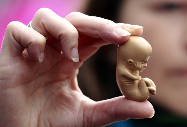
Researchers have come up with a new 3D model of human embryonic development over the first two months of pregnancy. Known as the 3D Atlas and Database of Human Embryology, it constitutes an important new resource for the scientific community and may shed a light on why congenital defects sometimes develop.
Current textbooks on early human development are for the most part outdated, as they rely on old articles and information published decades ago.
Researcher Bernadette S. de Bakker, from the University of Amsterdam. told IBTimes UK: "It's fair to say that we currently know more about the Moon than about our own embryonic development.
"The current textbooks all show the same kind of images based on embryonic specimens from the 1930s. Subsequent books kept using the image, only updating new molecular information."
She and colleague Antoon F M Moorman, helped by biomedical students, used embryo samples to create a three-dimensional digital atlas and database spanning the first two months of human development.
In total 15,000 samples from the Carnegie Collection of human embryonic specimens were analysed and included in the Atlas. The work involved identifying and labelling about 150 organs and structures, establishing changes in the position of organs.
"The 3D Atlas and Database of Human Embryology is the first to present such a large amount of data based on such a large number of human embryos. With this atlas and database we are able to quantify very precisely embryonic growth and the growth and position of specific organs," de Bakker says.
How the model may be used
The model will greatly help scientists working on early human development and development-related diseases. For instance, it solves a number of questions regarding kidney and gonad development in the human embryo.
It also confirms that there are important differences between human embryos and mice and chicks embryos, which are often used as models to study mammalian embryo development. Identifying these differences is crucial, so that they can be taken into account during this type of research.
"The atlas is useful because to study how congenital malformations appear it is important to know how normal human embryonic development occurs. Doctors will now be able to show pregnant women how and when congenital malformations occur. For scientists who study how toxic substances can affect embryonic development, this knowledge could help refine the chick and mice models they work with." concludes de Bakker.
© Copyright IBTimes 2024. All rights reserved.


