

3D-printed heart model helps surgeons save teenager from life-threatening aneurysm

Doctors at the Children's Hospital of Michigan in Detroit, US, have made a cardiovascular breakthrough by using a 3D-printed heart model to save the life of a teenager with a large, complex aneurysm in less than 24 hours and without surgery.
Because one of her brothers has a heart murmur, Ariana Smith, 17, an active teenager who plays volleyball and is a cheerleader, went for a routine electrocardiogram (EKG) in November 2014 to check to see if there were any problems with the electrical activity in her heart.
The test found that there might be potential abnormalities in her aorta (the main artery of the body), and so she was sent to the Children's Hospital of Michigan to have a cardiac catheterisation – a routine procedure whereby a tube is placed into the blood vessel so that doctors can see and properly diagnose the condition.
The doctors found that there was a huge aortic aneurysm with a tortuous aorta, i.e. an aorta that has a distorted shape. The condition does not produce any symptoms, but if the aneurysm were to rupture, it would prove deadly.
"We were devastated to hear this news. Ariana had no symptoms. I understand an aneurysm is considered a silent killer. We hoped and prayed that everything would turn out ok," said Ariana's mother Jacqueline Foster.
Pioneering medical procedure using stents
The heart specialists at the hospital considered treating Ariana using covered stents, which expand the artery and block the aneurysm, preventing it from rupturing. This method is currently being researched in an FDA-approved research study called the COAST II trial (Coarctation of the Aorta Stent Trial) and only a few hospitals in the US are able to offer this treatment.
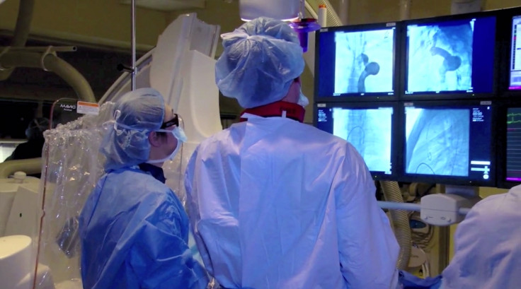
However, the procedure is very delicate and causes major complications if it is not performed properly. So the cardiologists turned to computed tomography to obtain medical image data of Ariana's heart, which was then passed to 3D-printing service provider Materialise.
Materialise fed the CT scan data into its Mimics Innovation Suite software and HeartPrint service, and was then able to create a 3D-printed life-like model of her heart, which the surgeons used to pinpoint the exact placement of the stent, to plan the treatment.
Using 3D printing to pin-point the stent placement
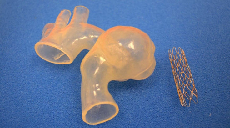
"Using the 3D-printed model of Ariana's aorta, we performed a 'practice-run' or simulation in the cath lab, where we actually placed a stent into the model," said Dr Daniel Turner of the Children's Hospital of Michigan.
"This allowed us to precisely plan the procedure and see how the stent responded in her unique and tortuous anatomy. Then, when we performed Ariana's actual procedure, we had a good idea of how it was going to go."
Usually, an aneurysm of this magnitude would require surgery, which is potentially risky and takes a longer time to recover from, but using the stents method, the doctors were able to treat her quickly and she was released from hospital within a day.
"We anticipate that Ariana will not require surgery to treat this condition. Dr Kobayashi will follow her closely in the office. Most importantly, this experience will allow us to treat future patients more safely with the use of 3D printing technology. This is only the beginning," said Turner.
© Copyright IBTimes 2024. All rights reserved.
-

Football Clubs With The Most Arrests And Bans in 2022-23
-

Oil Prices Slip As Investors Eye Israel's Response To Iran Strike
-
Billionaire Declared Missing And Dead In 2018 Is 'Possibly Living In Moscow With Mistress'
-

Wendy's Sued For $20M After 11-Year-Old Gets Brain and Kidney Damage From 'Dirty Meal'


