

Scientists Recreate Images of 300-Million-Year-Old Insects
One of the insects is a predecessor to the cockroach
Manchester University scientists have constructed 3D images of two 305-million-year-old juvenile insects using high resolution CT scan technology.
In a paper published on the peer reviewed scientific journal, PLOS ONE, the researchers said they were able to create 2,000 cross-section slices by placing fossils under the CT scanner and taking over 3,000 X rays from different angles.
These slices were further used to create three dimensional reconstructions of the insects, helping to learn about their lifestyle, biology and food habits.
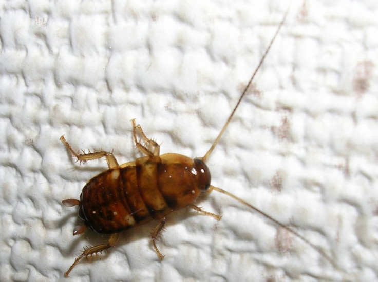
One of the two insects is an early predecessor of cockroach and is one of the best examples of the insect from the period ever seen by scientists, said a release from the university.
The other, which is extinct today, has a number of sharp spines.
Both are members of the subdivision Polyneoptera, which include insects like roaches, mantises, crickets, grasshoppers and earwigs.
Analysing the exact relationship of these creatures is difficult as insects tend to evolve a lot in appearance during their development.
"Around this time a number of early 'amphibians' were insectivores - they lived by eating a lot of insects," said Dr Russell Garwood, researcher at the university's School of Materials.
"The spiney creature was a sitting duck, as it couldn't fly, so the spines probably made it less palatable. It is bizarre - as far as we're aware, quite unlike any members of the Polyneoptera alive today," he added.
Garwood hopes that research such as this will help understand more about insects, especially their development and habitat. He said this was the first step and that he will be spending the next few years doing further studies on other fossil insects to build on this work.
Professor Philip Withers, co-author on the paper, said: "I am very excited by our fossil work which is providing unique information in 3D."
© Copyright IBTimes 2024. All rights reserved.


