This ultra-fast microscope captures 3D images of organisms in real time
The instrument can be used to monitor cellular changes in living organisms as they develop.
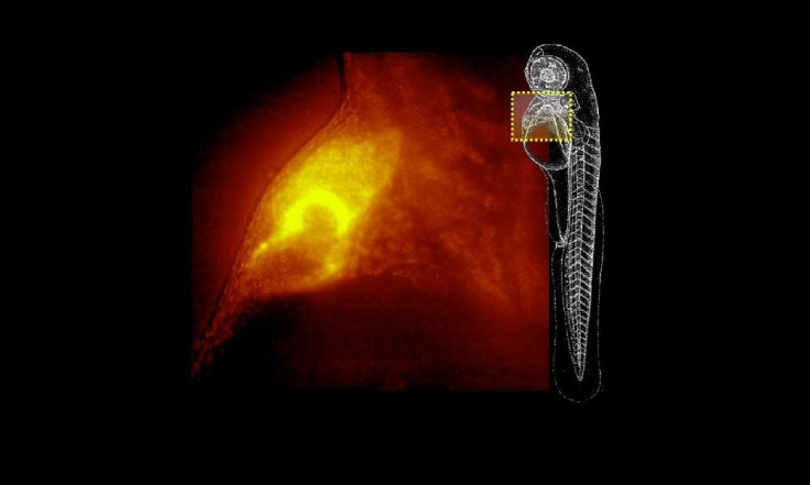
Scientists have developed a microscope that captures 3D, cellular-level images of living organisms in real time.
Dubbed QIs-scope, the new instrument uses a set of lasers, motors, cameras, and filters coordinated with sophisticated software. It takes up to 200 images every second, measuring a specimen from different angles and generating a 3D image. The image-capture velocity of this microscope goes far beyond the capability of modern confocal instruments that capture just around five images per second.
QIs-scope comes with a set of four lasers, each of which emits a flat beam of light and allows for marking cells or molecular processes with different colours. This helps with the monitoring of up to six different cells or different cell types in the same specimen.
"Our goal is for the QIs-scope to be easy to use with intuitive software, so that the user can see the specimen and choose where to make the scans, choose the excitation colors and generate a three-dimensional image with as many colors as were chosen," said Jorge Ripoll co-founder of 4D Nature – the company producing the new microscope.
The development of this instrument marks the latest step in step in confocal microscopy, and could soon help with biomedical researches or clinical diagnosis procedures. The images can also provide a detailed insight into the changes that occur during the development of a tissue or the internal functioning of organs, making it possible for the researchers to monitor animals as they develop.
"We can see how the heart of a zebrafish beats and make a 3D- reconstruction of its beat," Ripoll added. "It can be used for many studies related to cardiovascular illnesses, and to better understand how the heart functions."
According to a spokesperson of 4D Nature, "Right now there's no company that offers a team with similar characteristics. Other teams are ten times slower and cannot combine several angular measures in large samples".
© Copyright IBTimes 2024. All rights reserved.
-

Uber Eats Starts Robot Deliveries In Tokyo
-
Former "The Mandalorian" Actress Gina Carano Heaps Praise On Elon Musk, Compares Him To Batman
-

Quebec Approves X Gender Marker For Trans And Non-Binary People On Provincial Cards
-

Elon Musk Calls £4.42B Lawyer Fees 'Criminal' In Tesla Pay Dispute
-

France Enshrines Abortion As Constitutional Right In World First
-

Netizens Air Looting Concerns After Nike Announces First US 'World Of Flight' In Philadelphia
-

Copilot Offers Controversial Responses On Teaching Sex, DEI, LGBTQ Topics In Preschool