

Ultra small bacteria that may define new lower size limit of life detected
The smallest bacteria, which are believed to be as small as a living cell can get, have been detected in images taken by researchers using an electron microscope.
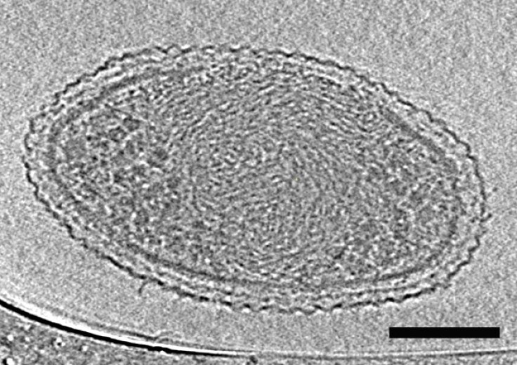
They are so small that 150,000 numbers can fit into the tip of a human hair. They are believed to be quite common.
The ultra-small bacteria isolated from groundwater belong to a phyla of microbes poorly understood.
Scientists from the US Department of Energy's Lawrence Berkeley National Laboratory and the University of California, Berkeley led the study which has been published in Nature Communications.
At an average volume of 0.009 cubic microns (one micron is one millionth of a metre), the cells are smaller than several estimates for the lower size limit of life required to sustain life.
The bacterial cells have densely packed spirals that are probably DNA, a very small number of ribosomes, hair-like appendages, and a low metabolism that may make them rely on other bacteria for sustenance.
Learning more about the organisms from these phyla could shed light on the role of microbes in the planet's climate, and other key processes, says the team.
"These newly described ultra-small bacteria are an example of a subset of the microbial life on earth that we know almost nothing about," says Jill Banfield, a Senior Faculty Scientist in Berkeley Lab's Earth Sciences Division and a UC Berkeley professor in the departments of Earth and Planetary Science and Environmental Science, Policy and Management.
While looking for small bacteria, the scientists filtered groundwater collected at Rifle, Colorado, through successively smaller filters, down to 0.2 microns, which is the size used to sterilise water.
But the final samples were not sterile, but steeped in microbial life.
The frozen samples were transported to Berkeley Lab, where the cells' size and internal structure was characterised using 2-D and 3-D cryogenic transmission electron microscopy.
The images also revealed dividing cells.
The bacteria's genomes were sequenced at the Joint Genome Institute and seen to be about one million base pairs in length.
The DNA studies revealed three phyla of bacteria in the samples.
"We don't know the function of half the genes we found in the organisms from these three phyla," says Banfield.
Recently, scientists estimated the cell volume of a marine bacterium at 0.013 cubic microns, but they used a technique that did not directly measure the cell diameter.
© Copyright IBTimes 2024. All rights reserved.


