

Folded structure of human brain determined by physical growth process rather than biology

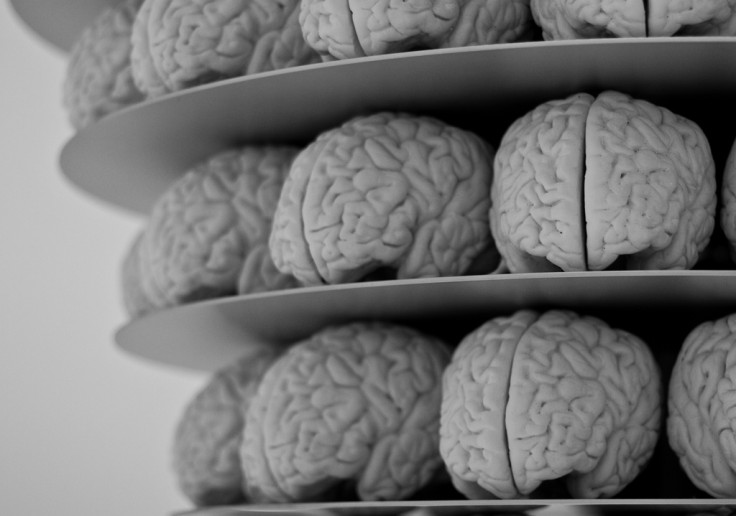
The folded structure of our brains appears to be the result of a physical growth process rather than a biological one. An international team of researchers used a 3D printing technique to mimic the growth of a human brain – and found the layers were created by mechanical compression forces.
Leading the study, published in Nature Physics, scientists from Harvard University in Cambridge, Massachusetts were looking at a model of brain formation first put forward more than 40 years ago. It said the brain's shape results from a growth process and has little to do with biology or chemistry of the brain.
Structure of the brain
Why human brains are folded is not known – and many studies into this area focus on genetics and the cellular regulation of growth. Evidence to support models regarding the structure is limited, however, because of the ethics surrounding experiments on human brains. In an accompanying News & Views article, Ellen Kuhl, from the the Departments of Mechanical Engineering and Bioengineering at Stanford University in California, notes that the average adult brain has a volume of 1,200cm cubed, a surface area of 2,000sq-cm and a cortical thickness of 2.5mm.
The process of folding is known as 'gyrification', and is thought to be a mechanism by which the number of cortical neurons is maximised while the distance between them is minimised. This process begins at around 23 weeks of gestation. Our brains continue grow until adulthood.
3D printing technique
In the study, researchers 3D-printed a model of a brain with soft gels based on an MRI of a foetal brain. This brain was made up of layers of different types of gel that were all designed to swell to different degrees when they were put into a solvent – mimicking human brain growth.
Their findings showed this growth "puts the outer layer into mechanical compression" and results in gyri and sulci – which create the folded appearance of the brain – similar to those seen in foetal brains.
"Starting with the same initial geometry, we also build numerical simulations of the brain modelled as a soft tissue with a growing cortex, and show that this also produces the characteristic patterns of convolutions over a realistic developmental course," they wrote. "All together, our results show that although many molecular determinants control the tangential expansion of the cortex, the size, shape, placement and orientation of the folds arise through iterations and variations of an elementary mechanical instability modulated by early foetal brain geometry."
Implications
The team say their findings have implications for diagnosing and treating several neurological disorders linked to malformation and brain size, as well as cell migration and cortex thickness. It also provides a template for physically based studies looking at the development of the brain surface, they note. Kuhl said, however, that while the findings are interesting, "the model can only predict the behaviour of simple, regular structures at the very onset of folding". She said re-creating the complicated and dynamic process of the emerging folds is challenging.
"Their simulations explain why folding always begins in weakly curved regions and why gyri and sulci align perpendicular to the direction of maximum compressive stress. Experiments with swelling brain models provide the essential missing link between modelling, experiment and simulation.
"However, a few limitations remain: the model is beautiful and simple, but it is limited to the initial folding of idealised structures; the experiment is useful for exploring instabilities beyond the onset of folding, but it is limited to moderate changes in volume."
She said further research could lead to a better understanding of the brain that can then be translated into medicine. For example, linking growth to neurological developments could result in scientists being able to trace specific brain functions back to changes in the brain's surface. "Making these connections can help us identify topological markers for the early diagnosis of autism, schizophrenia or Alzheimer's disease, and, ultimately, design more effective treatment strategies."
© Copyright IBTimes 2025. All rights reserved.
- MOST POPULAR IN Health


