Mini-brain in spinal cord vital for delicate motor skills like walking on ice
Balancing on a narrow ledge or slippery ice is possible thanks to the 'mini-brain' or cluster of neurons in the spinal cord that works to suitably adjust the muscles for the task.
Research done at Salk Institute saw scientists map in mice this mini-brain or neural circuitry of the spinal cord that deals with the sense of light touch and causes the body to reflexively make small adjustments to foot position.
This spinal circuit serves as a control centre for integrating motor commands from the brain with sensory information from the limbs.
Understanding the circuit better can help develop therapies for spinal cord injury and diseases that affect motor skills and balance.
"Our study opens what was essentially a black box, as up until now we didn't know how these signals are encoded or processed in the spinal cord. Moreover, it was unclear how this touch information was merged with other sensory information to control movement and posture," says Martyn Goulding, a Salk professor and senior author on the paper.
Balance sensors in our inner ear keep our heads level with the ground while sensors in our muscles and joints track the changing positions of our arms and legs. Multiple streams of signals from the newly identified light touch transmission pathway flow into the brain.
The eye uses neurons and light sensors to perform visual calculations before the information is sent to the visual centres in the brain.
But in the case of touch, the types of neurons involved and how they are wired together was not known.
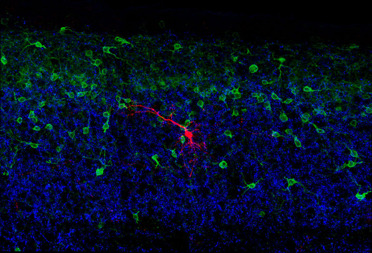
In their study, the Salk scientists traced nerve fibres that carry signals from the touch sensors to the spinal cord where they connect with a group of neurons known as RORα neurons. These serve as a critical link between the brain and the feet.
The RORα neurons not only receive signals from the brain and the light touch sensors, but also directly connect with neurons in the ventral spinal cord that control movement.
Thus, they are at the centre of a "mini-brain" in the spinal cord that integrates signals from the brain with sensory signals to make sure the limbs move correctly.
Mice with disabled RORα neurons were substantially less sensitive to movement across the surface of the skin or to a sticky piece of tape placed on their feet but could still walk.
However, they struggled to walk across a narrow beam, performing more clumsily than animals with intact RORα neurons.
"We think these neurons are responsible for combining all of this information to tell the feet how to move," says Steeve Bourane, a postdoctoral researcher in Goulding's lab and first author on the new paper.
"If you stand on a slippery surface for a long time, you'll notice your calf muscles get stiff, but you may not have noticed you were using them. Your body is on autopilot, constantly making subtle corrections while freeing you to attend to other higher-level tasks."
The findings are published in Cell.
© Copyright IBTimes 2025. All rights reserved.
- MOST POPULAR IN Science