Transplanted neurons act like the normal cells lost to brain damage
Transplanted embryonic neurons represent hope for many patients with brain diseases or head injuries.

Transplanted embryonic neurons may help people who have have suffered traumatic damage to their brains, as these cells quickly rebuild neural circuits and restore function in animal models, scientists have shown. A large number of patients, including those diagnosed with diseases such as Parkinson's or those who have suffered strokes or brain damage following a head injury, could potentially benefit from this technique.
In mammals, the adult brain has a very limited capacity to compensate for a loss of neurons linked to a traumatic brain injury or an illness. Transplanted embryonic neurons have in the past been shown to survive in the brains of newborn mice, raising the hopes of using neuronal transplantation to treat a range of neurological diseases. In particular, neural transplantation has also emerged in different clinical studies as a possible therapy for Parkinson's disease.
The problem is that even though transplantation aims to replace lost neurons, the extent to which new neurons can integrate into existing circuits is not known.
"Up to now, we didn't know to what extent transplanted neurons could take over the function of degenerated neurons in a given brain region or if they could receive the appropriate brain input", Magdalena Götz, author of the study published in Nature, told IBTimes UK.
Looking and acting like classic neurons
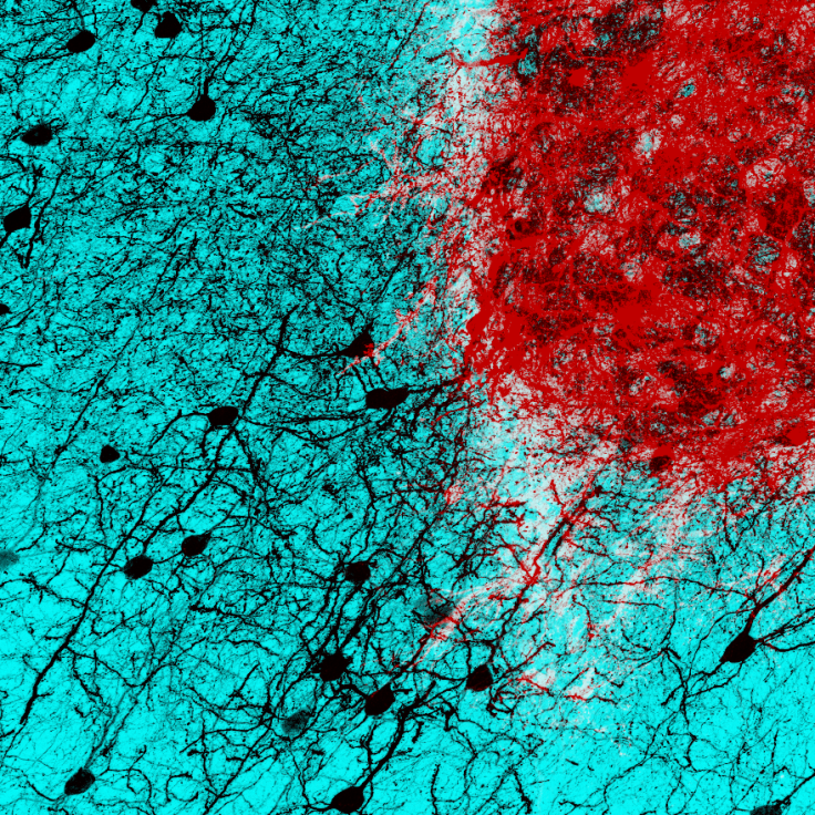
With her team, from the Ludwig Maximilians University of Munich, she used a sophisticated imaging technique known as chronic in vivo two-photon imaging to examine the fate of embryonic neurons, which has been transplanted into the damaged visual cortex of mice.
The researchers discovered that the transplanted neurons matured quickly, achieving adult like densities of dendritic spines and axonal boutons within four to eight weeks – they looked very much like the neuronal cells normally seen in the upper layers of the visual cortex in the absence of damage.
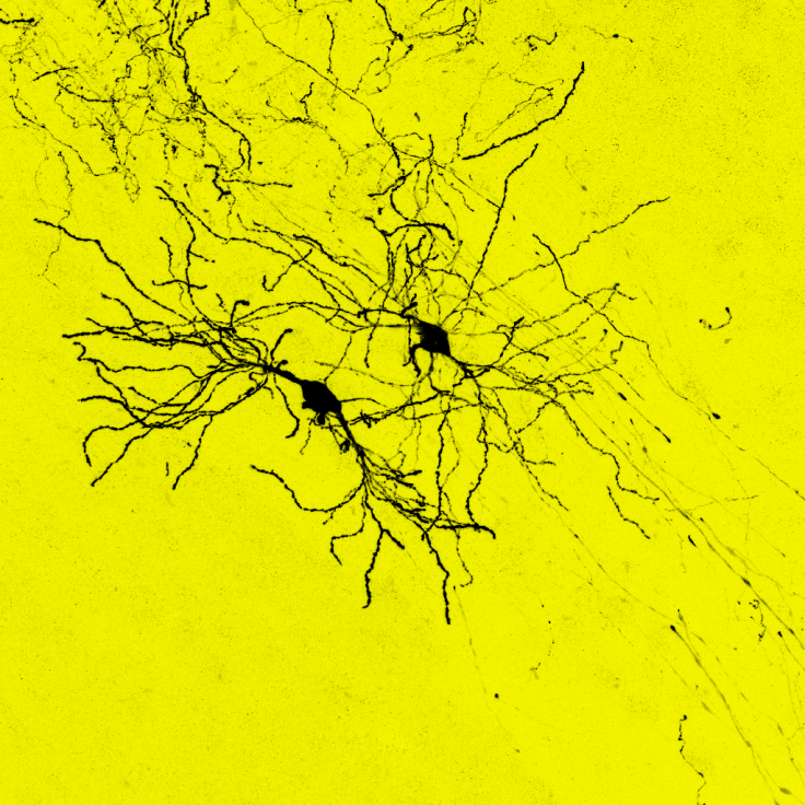
Even more important, these transplanted neurons were able to receive input from the rest of the brain, as they formed connections with host brain cells. This allowed them to receive electrical signals from other areas of the brain and to respond normally to visual stimuli. These transplanted neurons thus served the same function as the original neurons that had been damaged, essentially acting like them.
Further research has already began to follow the neurons in the damaged brains of mice models for other diseases. "This is just a proof of principle study, we will now study the way the transplanted neurons act in animal models of stroke or traumatic brain injury, which in the future may be more relevant to clinical models," Götz say.
© Copyright IBTimes 2025. All rights reserved.
- MOST POPULAR IN Health