What does the inside of a rock look like? Scientists can now give you a tour
Geologists used a camera, a grinder and AI to make a 3D model of a rock.
Using a high-resolution camera, an industrial grinder and the help of a custom-made neural network, a duo of geologists from Princeton University have devised a way to make a 3D representation of rocks that have fossils embedded in them.
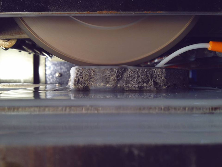
A release put out by the university details how this process works. A sample that needs to be studied is placed in the Princeton Grinding Imaging and Reconstruction Instrument (GIRI) and it is then ground out, layer by layer. However, these layers are unique since the instrument makes cuts in such a way that a layer can be as thin as 1 micron, noted the report.
As each layer is ground out, a high-resolution camera takes a picture of the newly exposed surface. A software then stitches together a detailed 3D image of the fossil that the rock has embedded within it. A sample that is about an inch thick takes around a day-and-a-half to complete, with each slice and image taking approximately 90 seconds.
GIRI was envisioned and developed by geologists Akshay Mehra and Adam Maloof at Princeton, where they have used it to create a 3D render of a 545 million-year-old Cloudina sample.
Cloudina, noted the report, is often considered to be one of the earliest forms of life that evolved the ability to "biomineralise". That means it was able to build a shell or bones, and not just soft tissue, for itself. "I thought going in, we would learn all sorts about this amazing first biomineraliser and first reef builder, but Cloudina turned out to be more like a reef dweller," said Maloof.
This finding can be considered a breakthrough because until GIRI was built, it was almost impossible to study Cloudina. The organism, now extinct, had shells and bodies that researchers are not been able to physically extract from the limestone it is embedded in because they are too delicate. Also, the shells of this pre-historic creature was identical in chemical makeup to limestone, which meant that X-rays were also useless.
Using GIRI, however, a high-resolution, detailed 3D model of a small sample was created, as seen in the video. The neural network which was used was trained to ignore rock and only image the Cloudina. So what is seen in the image is only the cluster of worm-like creatures.
However, to attain this level of resolution, any sample that has to be studied will be completely destroyed in the process.
"It's destructive of course, that's the disadvantage, but what's so nice is that you get to see photographs and make direct observations," said Maloof. "That's what's been so life-changing to me – I love that it's not a model. You can just see it. On any given slice, if you find something great, you can just find the slice and say, 'What did it look like?' ...We're on a virtual tour inside, rather than looking at waveforms and trying to interpret them."
While this method might seem like a waste of precious samples, the geologists obviously feel otherwise. There is no other way to effectively see inside a sample and not destroy it.
One method that was used before was to dissolve the surrounding material. However, Maloof said that the earlier process was not very effective.
"You lose all the in-situ information. You don't know how the fossils or other embedded objects grew. They have no relationship to each other. And you don't know how they are related to perhaps smaller or less resilient parts. You may preferentially dissolve the ornamentation or other key details," he explained.
- MOST READ