Brazilian Neurosurgeons Using 3D-Printed Brain Replicas to Save Children

While much has been written about doctors in Europe and China starting to use 3D-printing technology in medical applications, use of the technology is also on the rise in South America.
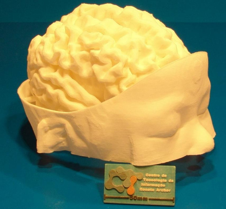
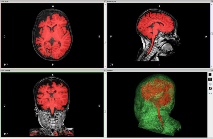
Recently, neurosurgeons at the University of São Paulo's medical school in the city of Ribeirão Preto in São Paulo, Brazil used 3D-printing to create a model of an infant's brain from a CT scan, in order to successfully treat a baby born with Sturge-Weber syndrome, a rare congenital neurological and skin disorder that severely affects a child's development.
The condition causes seizures, tumours, cerebral malformation (where the brain's arteries and veins do not connect properly as the brain develops), muscle weakness, glaucoma (a form of nerve damage which affects vision) and mental retardation, as well as unsightly birthmarks on the face from malformed blood vessels under the skin.
Physical therapy and anti-convulsant medications are usually used to treat the disorder, but in severe cases, doctors might choose to operate in order to disconnect or even "switch off" the affected part of the brain to prevent the life-threatening seizures.
The 3D-printed model of the infant's brain was used as an essential reference by Dr Hélio Rubens Machado at the University of São Paulo before and during the incredibly complex surgery, using technology provided by the Center for Information Technology Renato Archer (CTI).
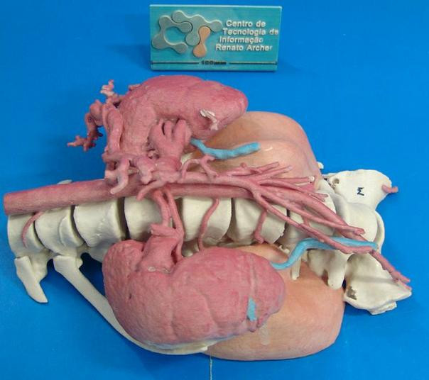
CTI has been making 3D-printed models for medical use for several years, such as a full colour model of a 12-year-old girl's spine, featuring a large tumour surrounding kidneys, arteries and the lungs, which was used to help prepare surgeons in removing it.
"CTI is the IT research centre from the Science and Technology Ministry of Brazil, developing and integrating solutions for better surgical planning," Jorge Vicente Lopes da Silva, head of the three-dimensional technologies division at CTI, told 3DPrint.com.
"We have many other applications [for 3D-printing] like Egyptology and palaeontology [and] we developed [the] experimental technology for about 3,500 cases with more than 100 hospitals in Brazil and some from Latin America."
Elsewhere in the world, Spanish doctors have used 3D-printing to remove a previously inoperable tumour from a child, while hospitals in Italy and the UK are using the technology to repair complex bone fractures and test out implants for facial reconstruction surgery.
In the Netherlands and China, surgeons are using 3D-printing in pioneering surgeries, including replacing an entire skull, and replacing a damaged vertebra.
© Copyright IBTimes 2025. All rights reserved.
- MOST POPULAR IN Technology