Brain created in a dish allows scientists to watch neural activity for the first time
The research could shed a light on a number of neurodevelopmental diseases.

Scientists have successfully assembled working human brain circuits in a dish. Their model could shed light on how a number of neurodevelopmental diseases arise, including rare forms of autism.
In contrast to research conducted in other fields of medicine, psychiatric research is often limited by poor methods and difficulty accessing brain tissues.
Although functional MRIs have greatly improved researchers' ability to examine the brain, these images do not represent its cellular morphology in all its details.
Post-mortem studies can help, but they are in no way sufficient to understand how the live brain works.
In recent years, pluripotent stem cells, which can differentiate into any adult cell types, have given scientists new hope. They have begun to study the developing human brain using neurons derived from these stem cells.
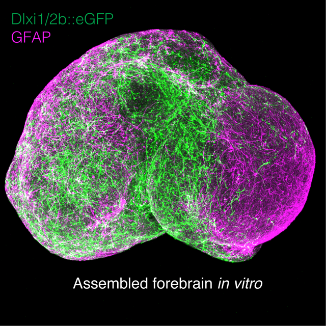
In a study now published in the journal Nature, a team has gone further, recreating the interactions between two types of brain cells crucial to the development of the human cerebral cortex.
"We have been making neurons in a dish for a few years now. If you look at these neurons under the microscope, you will see interesting things, but what you see won't be entirely 'brain-like'. The brain is a much more complex structure with different cell types positioned next to one another and communicating between each other. We wanted to go beyond simple models to study interactions between neurons," lead author Sergiu Pașca, from Stanford University, told IBTimes UK.
A rare form of autism
When the brain develops in the foetus, neurons known as GABAergic neurons migrate from deep tissues of the brain to integrate with glutamatergic neurons in the cerebral cortex. Scientists think that if something goes wrong with this migration and with the way these cells interact, this may lead to psychiatric diseases.
The researchers created two "bowls of neurons" out of pluripotent stem cells – one with GABAergic neurons and one with glutamatergic neurons.
They assembled them in a dish and observed how they fused over a number of weeks. They noted that the GABAergic neurons gradually started moving in a series of "jumps" towards the other neurons. The two cell types then started making connections.
Although this model is obviously not perfectly accurate – and not as complex as the brain itself – it is useful because it enables scientists to study cell migration and the development of functional human cortical circuits directly, in a dish.
The scientists put this model to good use to study a rare form of autism known as Timothy syndrome. They wanted to see if they could identify difference in brain cell migration when compared to healthy patients' cells.
Taking stem cells from patients with this disease, and turning them into brain cells, they were able to show with their model that GABAergic neurons' migration differed a little.
"In patients with Timothy syndrome, GABAergic neurons moved more often but were less efficient in their 'jumps'. We don't yet know what this means, but it does tell us that there might be something going on there which may explain the emergence of neurodevelopmental disorders – and that we may be able to use this system to study these processes that happen during foetal development," Pașca pointed out.
In a separate development, scientists from Harvard University published another study in the journal Nature describing how they managed to create "brain-like" clusters of cells in the lab, which may be kept in culture for over nine months, providing a window to analyse relatively late events of neuronal maturation.
© Copyright IBTimes 2025. All rights reserved.
- MOST POPULAR IN Health