UK Surgeons Develop New 3D Mapping Technique for Heart Rhythm Ops
Pioneering technique gives surgeons live images of 'short-circuits' in heart muscle
UK doctors have used a pioneering new technique to create 3D images of the human heart, to guide them during surgery to repair heart defects that cause dangerously irregular heartbeats.
Heart surgeons at St Thomas' Hospital and researchers at King's College have worked together to develop a brand-new procedure, that uses images taken by an MRI scanner to build a 3D image of the heart. Surgeons then use the images to find the areas of the heart that are causing the irregular heartbeats, and treat them.
Previously doctors could only use X-rays to take scans, which produced less reliable snapshots of the heart, but no live, real-time images. Using X-rays, doctors could only take a limited number of images because of the risk of exposing people to potentially harmful radiation, and the quality of the images were limited.
The new technique has the advantage of avoiding radiation exposure, and producing better quality, realtime images.
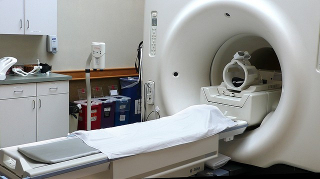
During the operation surgeons use an MRI machine to scan the upper half of patients' bodies to produce live images of the heart, which surgeons can use to detect which cells in the heart are "short-circuiting" – firing incorrectly and causing an irregular heartbeat, or "heart flutter".
Then – while the patient is still inside the MRI scanner – surgeons insert a catheter into the patient's groin, feed specially-designed instruments through to the blood vessels in the heart, and then use the tools to find and destroy the faulty cells: a technique called ablation.
Two men and a woman have already been successfully treated using the new techniques at St Thomas' Hospital in London. If left untreated, irregular heartbeats can wear out the heart muscle and stop the heart from working properly.
Professor Reza Razavi, head of imaging sciences at King's College London, who jointly developed the new type of operation with Professor Mark O'Neill, presented the results from the trials in three patients at a medical conference last week.
He told the Sunday Times: "The MRI lets us see a 3D image of the heart on a screen, to insert two catheters. We used them first to map out the heart and assess the electrical impulses and then to work out which tissues need ablating to block the short-circuit and so halt the flutter."
© Copyright IBTimes 2025. All rights reserved.
- MOST POPULAR IN Health