X-Ray Imaging Technique Allows Scientists to See Cystic Fibrosis Treatments 'Live'

A breakthrough technique allowing scientists to see how cystic fibrosis treatments are working "live" has been developed.
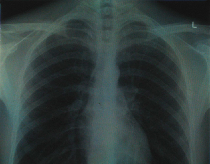
Researchers from Monash University in Melbourne, Australia, have developed an x-ray imaging system allowing them to watch treatments working non-invasively.
Published in the American Journal of Respiratory and Critical Care Medicine, the scientists say their system will allow doctors and researchers to measure how effective treatments are, meaning they can progress new treatments to the clinic faster.
It works by looking at soft tissue structures like the brain, airways and lungs, which are currently invisible in conventional x-ray machines.
Kaye Morgan, lead researcher on the paper, said: "At the moment we typically need to wait for a cystic fibrosis treatment to have an effect on lung health, measured by either a lung CT scan or breath measurement, to see how effective that treatment is.
"However the new imaging method allows us for the first time to non-invasively see how the treatment is working 'live' on the airway surface."
Cystic fibrosis is a genetic condition where the sufferer's lungs and digestive system become clogged with thick sticky mucus. The life expectancy of a person with cystic fibrosis who survives to adulthood is 37.
At present it can take months to measure how effective treatment is. With the new imaging system, it would be instantaneous.
"Because we will be able to see how effectively treatments are working straight away, we'll be able to develop new treatments a lot more quickly, and help better treat people with cystic fibrosis," Morgan said.
© Copyright IBTimes 2025. All rights reserved.
- MOST POPULAR IN Health