The winners of the annual Nikon Small World Competition have been revealed. Now in its 43rd year, the contest combines art and science as it recognises the best photos taken under a microscope. This year's top images include a ghoulish tapeworm, a bejewelled bee, Pac-Man algae and weevil porn.
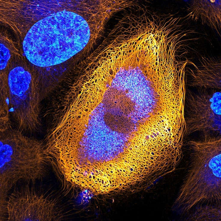
Dr Bram van den Broek of The NetherlandsCancer Institute (NKI) takes first place for his photo of a skin cell expressing an excessive amount of keratin. He came across this peculiar but beautiful skin cell while researching the dynamics of keratin filaments with Andriy Volkov, a student in the Cell Biophysics group led by Professor Kees Jalink.
"There are more than 50 different keratin proteins known in humans. The expression patterns of keratin are often abnormal in skin tumor cells, and it is thus widely used as tumour marker in cancer diagnostics," said. Dr van den Broek. "By studying the ways different proteins like keratin dynamically change within a cell, we can better understand the progression of cancers and other diseases."
1st place: Dr. Bram van den Broek, Andriy Volkov, Dr. Kees Jalink, Dr. Nicole Schwarz & Dr. Reinhard Windoffer, The Netherlands Cancer Institute, BioImaging Facility & Department of Cell Biology, Amsterdam, The Netherlands. Immortalised human skin cells (HaCaT keratinocytes) expressing fluorescently tagged keratin. Confocal. 40x (objective lens magnification)Dr Bram van den Broek, Andriy Volkov, Dr Kees Jalink, Dr Nicole Schwarz & Dr Reinhard Windoffer
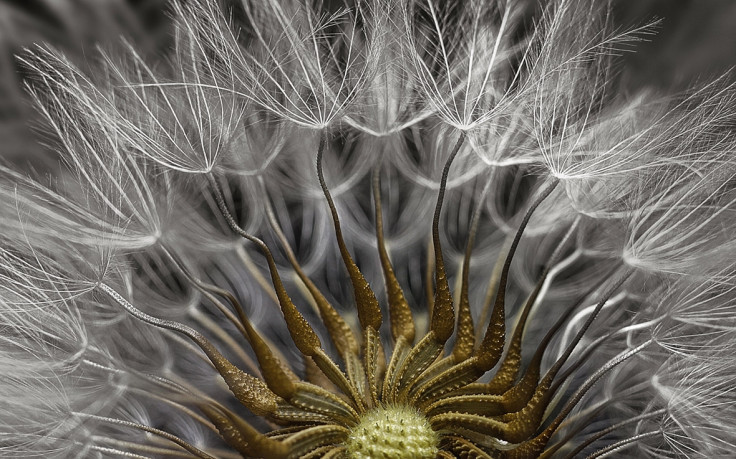
Dr Havi Sarfaty takes second place for his photo that captures a subject we see every day – the flowering head of a plant (Senecio vulgaris). Dr. Sarfaty is a veterinary ophthalmologist and has been taking photos through a microscope for about eight years – his interest sparked while performing eye surgeries
2nd Place: Dr. Havi Sarfaty, Eyecare Clinic, Yahud-Monoson, Israel. Senecio vulgaris (a flowering plant) seed head. Stereomicroscopy. 2xDr. Havi Sarfaty
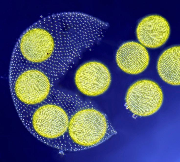
Although it looks like it came from a well-known vintage video game, Jean-Marc Babalian's third place image depicts a living volvox algae releasing its daughter colonies. Babalian has been taking photographs through the microscope for 30 years. He focuses his work on showing the fine details on ciliates and diatoms.
3rd Place: Jean-Marc Babalian, Nantes, France. Living Volvox algae releasing its daughter colonies. Differential Interference Contrast. 100xJean-Marc Babalian
IBTimes UK presents the rest of the top 20 images as chosen by the judges out of more than 2,000 entries from 88 countries around the world.
4th Place: Teresa Zgoda, Rochester Institute of Technology, Rochester, New York, USA. Taenia solium (tapeworm) everted scolex. 200xTeresa Zgoda5th Place: Dean Lerman, Netanya, Israel. Mould on a tomato. Reflected Light, Focus Stacking. 3.9xDean Lerman6th Place: Dr. David A. Johnston, University of Southampton/University Hospital Southampton, Biomedical Imaging Unit, Southampton, United Kingdom. Lily pollen. Confocal. 63x (objective lens magnification)Dr. David A. Johnston7th Place: Dr. Ryo Egawa, Nagoya University, Graduate School of Medicine, Nagoya, Japan. Individually labeled axons in an embryonic chick ciliary ganglion. Differential Interference Contrast. Confocal, Tissue Clearing, Brainbow (labelling technique). 30x (objective lens magnification)Dr. Ryo Egawa8th Place: Dr. Michael Perny, University of Bern, Institute for Infectious Diseases, Bern, Switzerland. Newborn rat cochlea with sensory hair cells (green) and spiral ganglion neurons (red). Confocal. 100xDr. Michael Perny9th Place: Catarina Moura, Dr. Sumeet Mahajan, Dr. Richard Oreffo & Dr. Rahul Tare, University of Southampton, Institute for Life Sciences, Southampton, United Kingdom. Growing cartilage-like tissue in the lab using bone stem cells (collagen fibres in green and fat deposits in red). Second Harmonic Generation (SHG) and Coherent Anti-Stokes Raman Scattering (CARS). 20x for collagen; 40x for fat depositsCatarina Moura, Dr. Sumeet Mahajan, Dr. Richard Oreffo & Dr. Rahul Tare10th Place: Dr. Csaba Pintér, University of Pannonia, Georgikon Faculty, Department of Plant Protection, Keszthely, Hungary. Phyllobius roboretanus (weevil). Stereomicroscopy. 80xDr. Csaba Pintér11th Place: Steven Simon, Simon Photography, Grand Prairie, Texas, USA. Plastic fracturing on credit card hologram. 10x (objective lens magnification)Steven Simon12th Place: Charles Krebs, Charles Krebs Photography, Issaquah, Washington, USA. Opiliones (daddy longlegs) eye. Reflected Light, Image Stacking. 20x (objective lens magnification)Charles Krebs13th Place: Levon Biss, Levon Biss Photography Ltd, Ramsbury, United Kingdom. Exaerete frontalis (orchid cuckoo bee) from the collections of the Oxford University Museum of Natural History. Reflected Light. 10x (objective lens magnification)Levon Biss14th Place: David Millard, Austin, Texas, USA. Common Mestra butterfly (Mestra amymone) eggs, laid on a leaf of Tragia sp. (Noseburn plant). Incident Illumination, Image Stacking. 7.5x (objective lens magnification)David Millard15th Place: Dr. Rick Adams, University of Northern Colorado, Department of Biological Sciences, Greeley, Colorado, USA. 3rd trimester fetus of Megachiroptera (fruit bat). Darkfield, Stereomicroscopy. 18xDr. Rick Adams16th Place: Marek Miś, Marek Miś Photography, Suwalki, Poland. Parus major (titmouse) down feather. Polarized Light, Darkfield. 25xMarek Miś17th Place: Harald K. Andersen, Steinberg, Norway. Dyed human hair. Darkfield. 40xHarald K. Andersen18th Place: Christian Gautier, Biosphoto, Le Mans, France. Synapta (sea-cucumber) skin. Polarised Light. 100xChristian Gautier19th Place: Dr. Dylan Burnette, Vanderbilt University School of Medicine, Department of Cell and Developmental Biology, Nashville, Tennessee, USA. Embryonic body wall from a developing Mus musculus (mouse). 100x (objective lens magnification)Dr. Dylan Burnette20th Place: Tracy Scott, Ithaca, New York, USA. Aspergillus flavus (fungus) and yeast colony from soil. Transmitted Light. 40xTracy Scott
Nikon Small World was founded in 1974 to recognise excellence in photography through the microscope. 2017 marks the 43rd year of the Nikon Small World Competition. This year is also the 100th anniversary of Nikon Instruments. Top images from the 2017 Nikon Small World Competition will be exhibited in a full-colour calendar and through a museum tour of the US and Canada. For additional information, visit www.nikonsmallworld.com.