Engineers develop hand-held pen-sized microscope to identify cancer cells outside the lab

Medical engineers from the University of Washington are developing a miniature, hand-held microscope that enables surgeons to distinguish between healthy cells and cancerous cells without needing to send samples to a laboratory, which could revolutionise cancer patient care.
It's all very well to send tissue samples off to a pathology lab to be analysed under a microscope in order to distinguish between normal and cancerous brain cells, but during a crucial operation when a surgeon has opened a patient's skull in order to remove a malignant brain tumour, there isn't time to wait for lab results. The surgeon needs to make sure that every single bit of cancerous material is removed from the brain, while ensuring that the healthy brain matter is protected, in order to reduce any chance of harm to brain from the operation.
"Surgeons don't have a very good way of knowing when they're done cutting out a tumour. They're using their sense of sight, their sense of touch, pre-operative images of the brain – and often times it's pretty subjective," said senior author Jonathan Liu, UW assistant professor of mechanical engineering. "Being able to zoom and see at the cellular level during the surgery would really help them to accurately differentiate between tumour and normal tissues and improve patient outcomes."
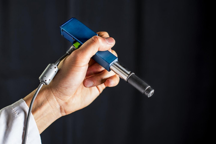
The miniature microscope is roughly the size of a pen and it uses dual-axis confocal microscopy to help surgeons see more clearly through opaque tissues, capturing details up to 0.5mm beneath the tissue surface, where some types of cancerous cells originate.
The device is also able to record and produce high resolution images of the tumour mass detected much faster than existing devices by using a technology called line scanning, where micro-electrical-mechanical mirrors direct an optical beam to scan tissue line by line to build up a picture.
Microscope produces images comparable with labs

At the moment, when doctors detect any suspicious moles on the body or legions in the mouth, they will usually cut them out and then send the tissue to a laboratory to be biopsied for cancer, and most of the time, these samples come back as benign.
Unfortunately, this process overloads pathology labs with too many tests to run and also subjects patients to an invasive procedure, so the miniature microscope could help doctors and dentists to better assess whether legions and moles are normal or suspicious, as the images that have been collected of mouse tissues are just as good as the images produced by a clinical pathology lab, which takes days.
The research, entitled "Miniature in vivo MEMS-based line-scanned dual-axis confocal microscope for point-of-care pathology" is published in the journal Biomedical Optics Express.
The researchers will next test out the miniature microscope as a cancer-screening tool in clinical trials in 2017, and hope that the invention can launched commercially for use in surgeries and other clinical procedures within the next two to four years.
"For brain tumour surgery, there are often cells left behind that are invisible to the neurosurgeon. This device will really be the first to let you identify these cells during the operation and determine exactly how much further you can reduce this residual," said project collaborator Nader Sanai, professor of neurosurgery at the Barrow Neurological Institute in Phoenix. "That's not possible to do today."
© Copyright IBTimes 2025. All rights reserved.
- MOST READ