UCLA's holographic microscope enables scientists to study spinning head of sperm in 3D
The microscope could be used to study and differentiate healthy sperm cells from defective ones.
A team from the University of California, Los Angeles (UCLA), has developed a 3D holographic microscope that, along with its image reconstruction algorithms, has made it possible to study the movement of sperm cells in precise detail.
The microscope is not only useful for observing sperm, but could also shed some new light on micro robotics. By mimicking the way sperm reacts and moves inside its environment could help in creating microscale robots, said Aydogan Ozcan, UCLA's Chancellor's Professor of Electrical and Computer Engineering and Bioengineering.
The computational imaging platform, or holographic microscope built by the team is composed of simple, inexpensive components, explains a publication by phys.org, making use of imaging sensors similar to the ones found in mobile phones and LEDs.
"Our holographic microscope replaces lenses with algorithms, and can image sperms over a sample volume that is approximately 100 times larger than that of standard optical microscopes," said Mustafa Daloglu, a UCLA doctoral student and first author of the study.
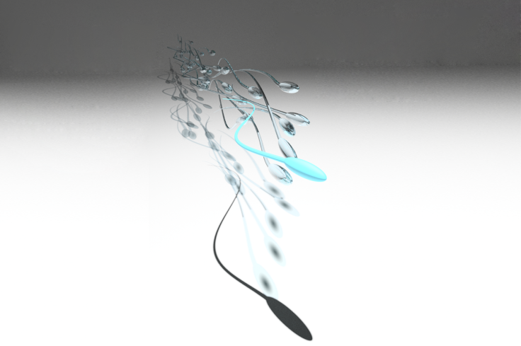
He added that this was the first time that the spinning sperm head and the freely beating patterns of the sperm cell's tail were reported in 3D. Traditional microscopes till now have been able to only observe sperm in a 2 dimensional view across a small volume and so were not able to capture its 3D movement.
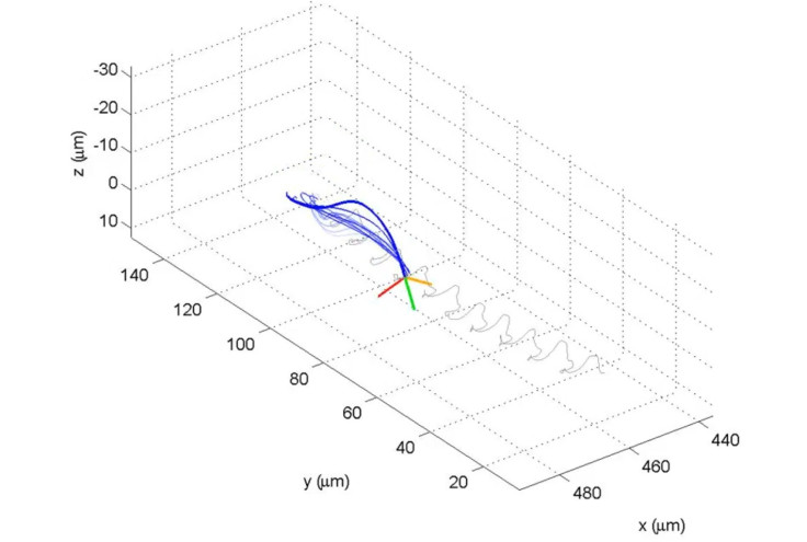
The sperm cell is placed in a "large observation chamber" which is around 30 microlitres in volume – 0.03 ml. This chamber is then placed on top of the image sensor and the LEDs are focussed in such a way that they cast two distinct shadows of the sperm swimming inside the chamber.
"Because of how the two LEDs are positioned, each individual sperm generates two separate shadows from different angles – each containing holographic information that is used to reconstruct a digital 3-D image of the sperm body," said Wei Luo, co-author of the paper.
© Copyright IBTimes 2025. All rights reserved.
- MOST READ